
Discover how our reliable treatments can safeguard your sight
Eye treatment options can be confusing. We’ve made the journey as straightforward as can be


Give us a call to understand how your insurance can help cover your treatment



Get a quick overview of everything you need to know about gaining visual freedom
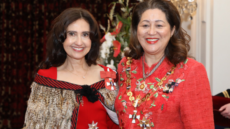
Celebrating Professor Dame Helen Danesh-Meyer
Professor Dame Helen Danesh-Meyer was formally recognised at an investiture ceremony in Wellington on [...]
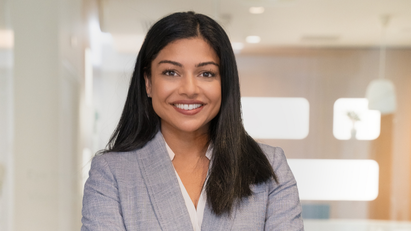
Dr Narme Deva joins ONZ Board
We’re incredibly proud to share that Dr Narme Deva has been voted onto the board of Ophthalmology New Zealand (ONZ) — a significant achievement [...]
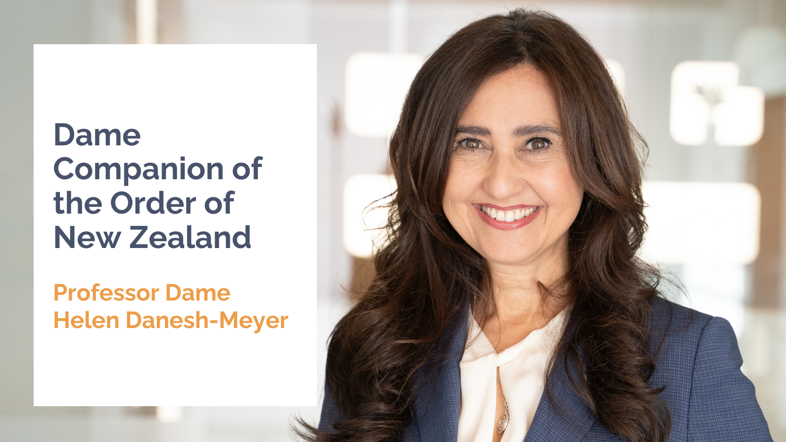
Damehood for leading ophthalmologist
Damehood for leading ophthalmologist Professor Helen Danesh-Meyer – the first ever female professor of [...]
Our industry-leading experts are here to guide you every step of the way
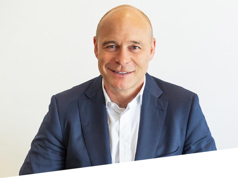
Dr Nick Mantell is a Refractive laser, Refractive Lens Exchange and Cataract Specialist. He has been working in private practice for 20 years.
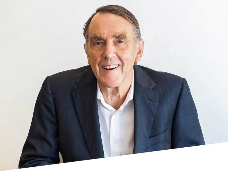
With four decades at the forefront of eye surgery in New Zealand, Dr Ring was an early exponent of modern multi-focal technology. He was also part of the esteemed small team that introduced laser surgery to the country.
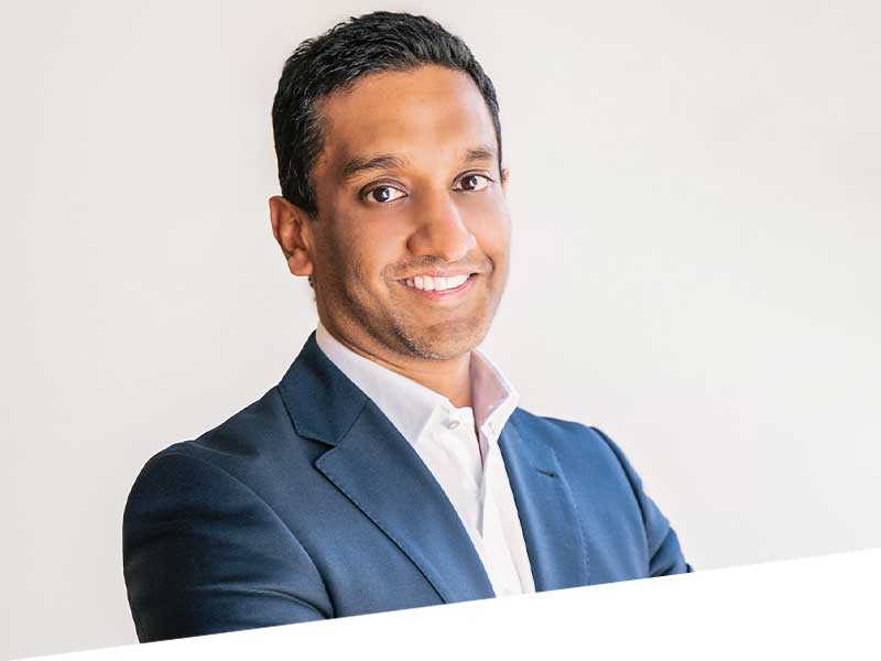
A cataract, corneal and laser surgery specialist Dr Subbiah has been with Eye institute since 2013. His easy manner and affable nature have found a natural home in New Zealand medicine.
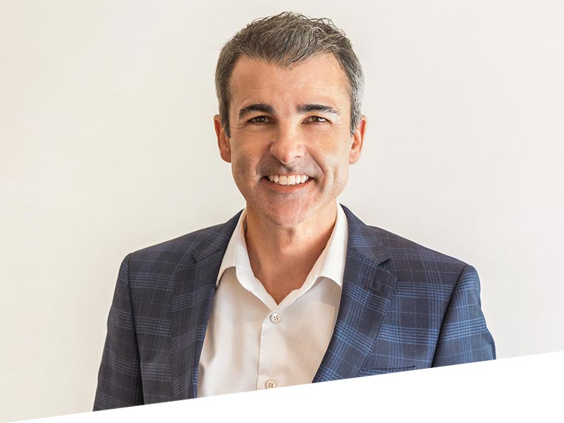
Dr Watson’s focus – pun intended – is the front of the eye. His combination of specialisations enables him to address the face, eyelids, tear drainage system, the surface of the eye and cornea together as a single functional unit to give the best possible outcome.
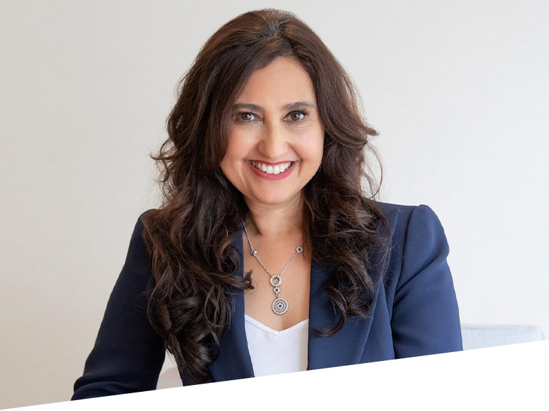
Professor Danesh-Meyer is an international authority on glaucoma and neuro-ophthalmology and one of the most highly regarded experts in her field.
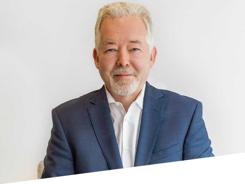
Professor Charles McGhee, a Consultant Ophthalmic Surgeon for 25 years, specialises in corneal diseases and cataract surgery.
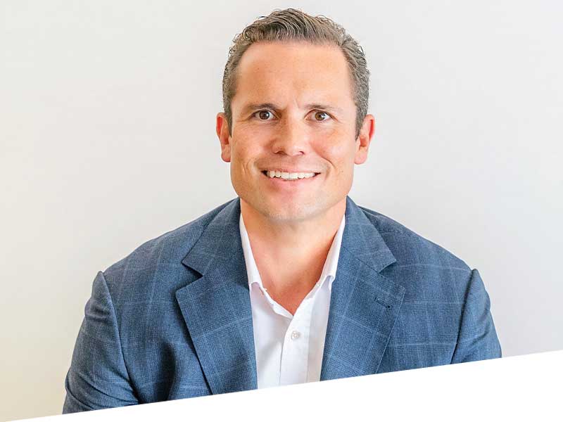
Will is an acknowledged expert in cataract and vitreoretinal surgery. He splits his time between the Eye Institute and works publicly at the Greenlane Clinical Centre.
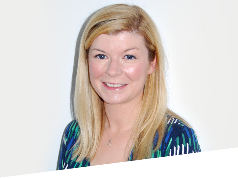
Dr Hill completed her MBBS at St Georges University of London in 2008, also obtaining an Intercalated BSc from King’s College University, London.
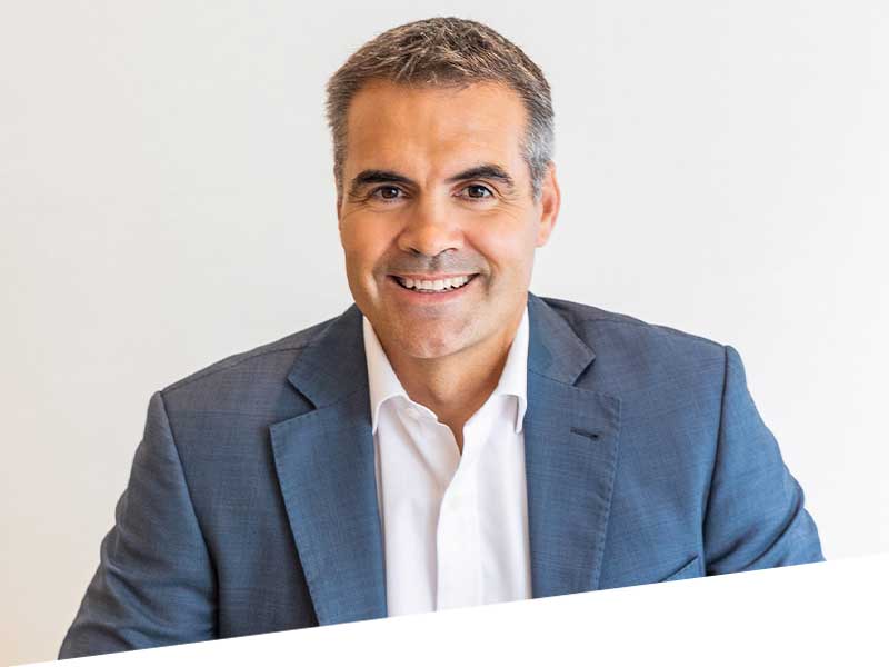
Dr Hadden specialises in vitreoretinal and cataract surgery, including the treatment of intraocular tumours. He has been at Eye Institute for 17 years.
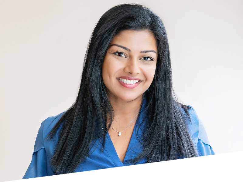
Dr Narme Deva is a key opinion leader for Medical retina and Uveitis in New Zealand and an expert cataract surgeon. She has a logical approach, an empathetic manner and a passion for the pursuit of excellence.
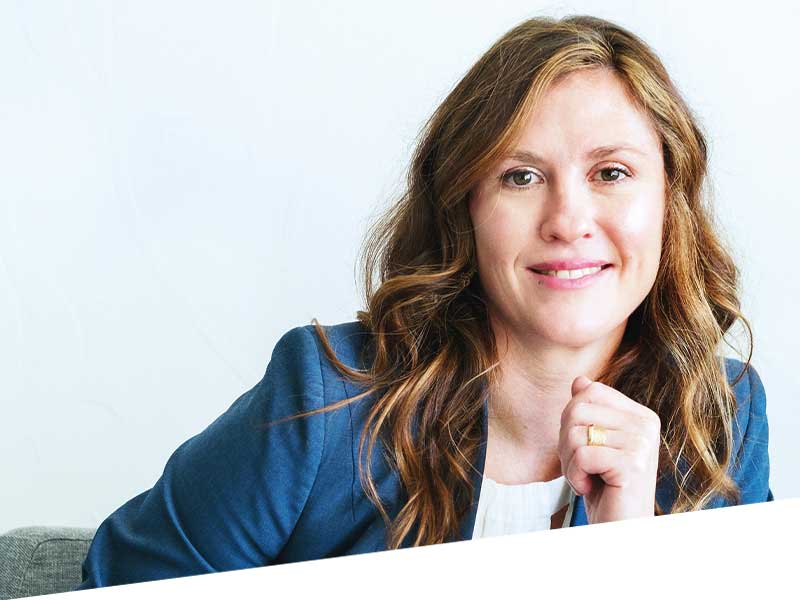
Dr Elizabeth Insull is a New Zealand and UK trained Oculoplastic and Ophthalmic surgeon based at Eye Institute Hawke’s Bay at Kaweka Hospital.
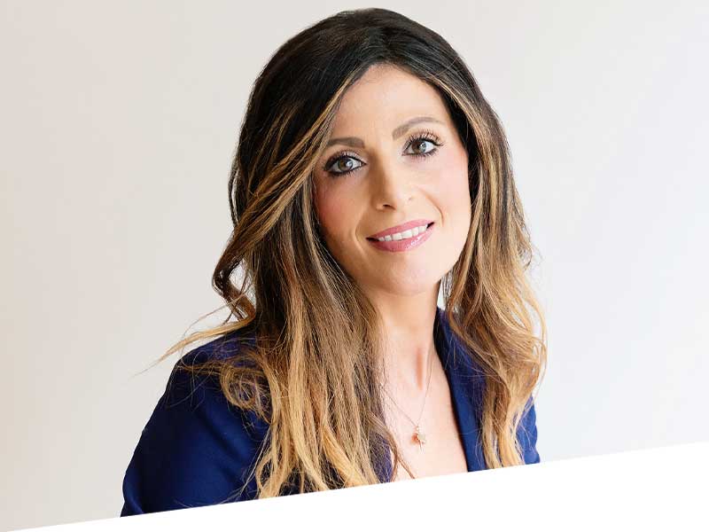
Dr Kaliopy Matheos is a New Zealand-trained Ophthalmologist specialising in Glaucoma & Anterior Segment Surgery at the University of Toronto in Canada.
As part of his ophthalmology specialty training, Dr Dunne gained valuable experience in major centres such as Auckland and Wellington.
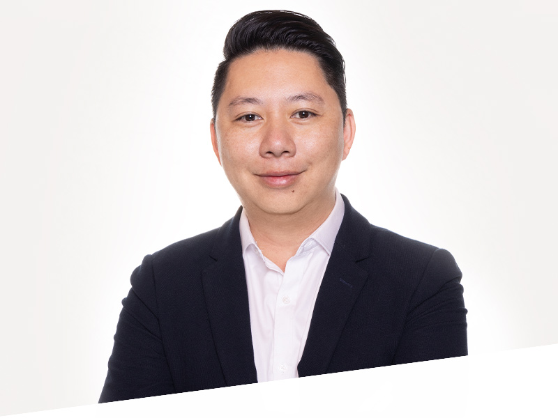
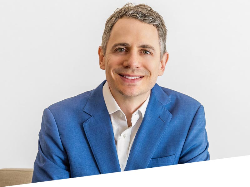
Dr. Jay Meyer is a specialist eye surgeon with expertise in cataracts, glaucoma, and corneal diseases and surgeries. He has a gentle demeanour and provides personalised care by tailoring treatments to each patient’s needs.
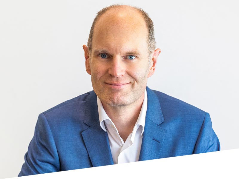
Dr Reeves has advanced training in the diagnosis and management of glaucoma. He offers a variety of laser and surgical treatments including minimally invasive glaucoma surgery.
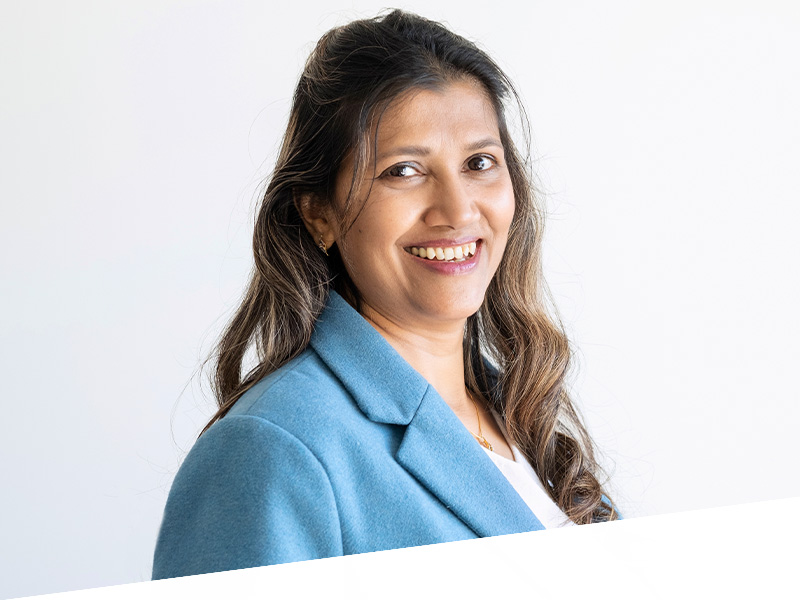
Dr Baswati Sahoo
Dr Baswati Sahoo is a cataract and glaucoma surgeon from India with over 15 years of experience in Ophthalmology.
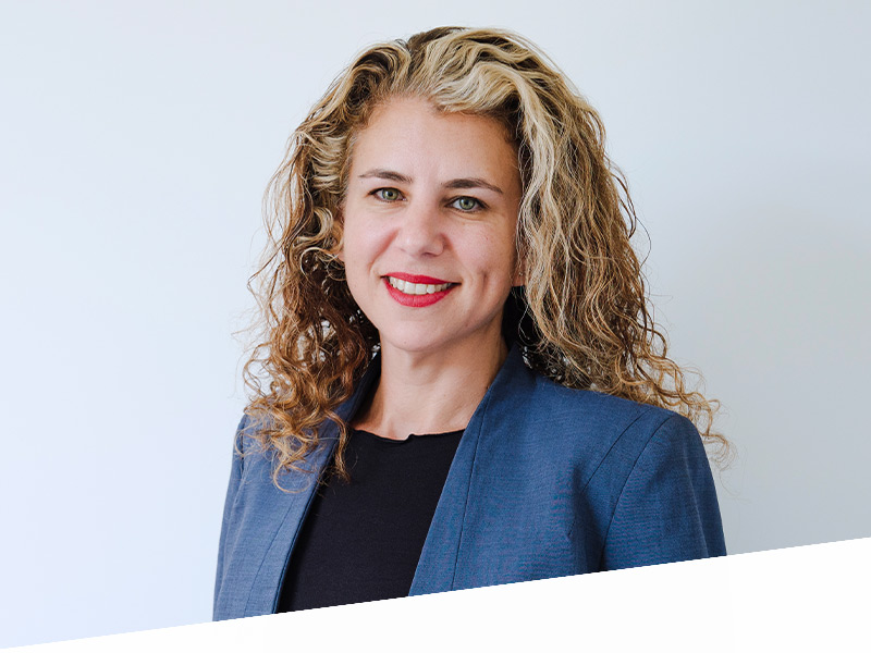
Dr Shira Sheen
Shira completed Ophthalmology training in Israel, under the supervision of world known Ophthalmologist, Professor Ehud Assia.
Dr Ammar Binsadiq
Dr Ammar Binsadiq is a New Zealand-trained ophthalmologist with subspeciality training in medical retinal, cataract and vitreoretinal surgery.
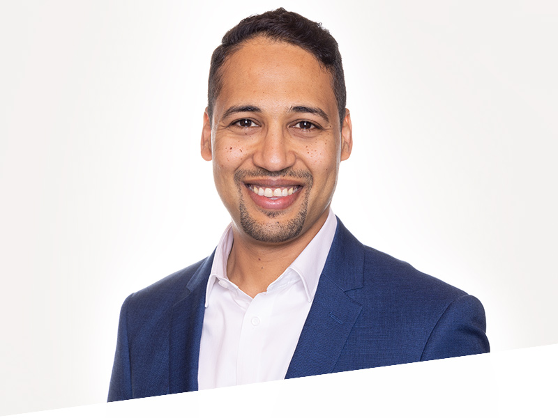
Dr Narme Deva is a key opinion leader for Medical retina and Uveitis in New Zealand and an expert cataract surgeon. She has a logical approach, an empathetic manner and a passion for the pursuit of excellence.
LEARN MORE
With four decades at the forefront of eye surgery in New Zealand, Dr Ring was an early exponent of modern multi-focal technology. He was also part of the esteemed small team that introduced laser surgery to the country.
LEARN MORE
A cataract, corneal and laser surgery specialist Dr Subbiah has been with Eye institute since 2013. His easy manner and affable nature have found a natural home in New Zealand medicine.
LEARN MORE
Dr Watson’s focus – pun intended – is the front of the eye. His combination of specialisations enables him to address the face, eyelids, tear drainage system, the surface of the eye and cornea together as a single functional unit to give the best possible outcome.
LEARN MORE
Professor Danesh-Meyer is an international authority on glaucoma and neuro-ophthalmology and one of the most highly regarded experts in her field.
LEARN MORE
Professor Charles McGhee, a Consultant Ophthalmic Surgeon for 25 years, specialises in corneal diseases and cataract surgery.
LEARN MORE
Will is an acknowledged expert in cataract and vitreoretinal surgery. He splits his time between the Eye Institute and works publicly at the Greenlane Clinical Centre.
LEARN MORE
Dr Hadden specialises in vitreoretinal and cataract surgery, including the treatment of intraocular tumours. He has been at Eye Institute for 17 years
LEARN MORE
Dr Elizabeth Insull is a New Zealand and UK trained Oculoplastic and Ophthalmic surgeon based at Eye Institute Hawke’s Bay at Kaweka Hospital.
LEARN MORE
Dr Kaliopy Matheos is a New Zealand-trained Ophthalmologist specialising in Glaucoma & Anterior Segment Surgery at the University of Toronto in Canada.
LEARN MORE
Dr. Jay J. Meyer is a specialist eye surgeon with expertise in cataracts, glaucoma, and corneal diseases and surgeries. He has a gentle demeanour and provides personalised care by tailoring treatments to each patient’s needs.
LEARN MORE
Dr Reeves has advanced training in the diagnosis and management of glaucoma. He offers a variety of laser and surgical treatments including minimally invasive glaucoma surgery.
LEARN MORE
Dr Nick Mantell is a Refractive laser, Refractive Lens Exchange and Cataract Specialist. He has been working in private practice for 20 years.
LEARN MORE
Dr Hill completed her MBBS at St Georges University of London in 2008, also obtaining an Intercalated BSc from King’s College University, London..
LEARN MORE
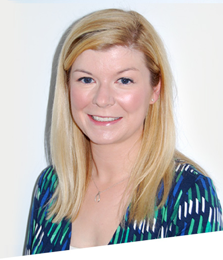
As part of his ophthalmology specialty training, Dr Dunne gained valuable experience in major centres such as Auckland and Wellington.
LEARN MORE
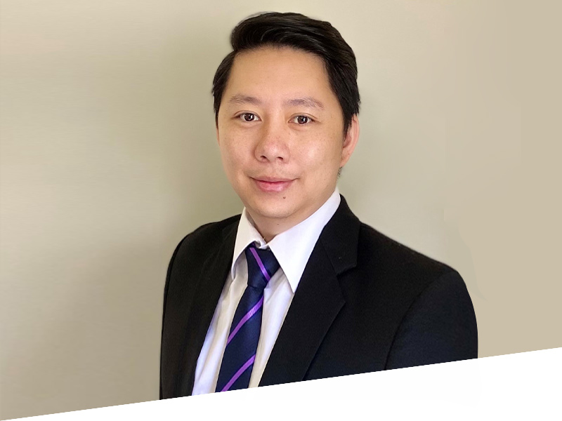
Dr Ammar Binsadiq is a New Zealand-trained ophthalmologist with subspeciality training in medical retinal, cataract and vitreoretinal surgery.
LEARN MORE
Dr Baswati Sahoo is a cataract and glaucoma surgeon from India with over 15 years of experience in Ophthalmology.
LEARN MORE
Dr Shira Sheen completed Ophthalmology training in Israel, under the supervision of world known Ophthalmologist, Professor Ehud Assia.
LEARN MORE
Dr Edward Hutchins specialises in Cataract, Corneal, Medical Retina & Vitreoretinal surgery.
LEARN MORE
Learn more about treating these corneal conditions from authoritative sources.
Note: These links will take you off our website